RADIOLOGY
(IMAGING SERVICES)
Radiology is a field of medicine that displays diseases using technologically advanced devices, makes a diagnosis and makes a differential diagnosis. Previously, this service, which could only be provided with X-ray devices, is now performed using a wide variety of advanced technical tools today. It can be said that radiology is the fastest-growing branch of medicine. With the rapid development of the disease, the chances of better treatment have been established by diagnosing the disease in more detail. For this reason, in order to treat a disease well, the necessity of conducting radiological examinations for that disease in detail with the radiological devices related to that disease is increasing day by day. But Radiological examinations should be performed as often as necessary and as much as necessary.
Our imaging devices;
– MRI Device (1.5 Tesla)
– Whole body computed tomography device
– Four-dimensional ultrasonography
– Three-dimensional Color doppler ultrasonography
– Mammography
– Digital (Computed) X-Ray
– Mobile (mobile) X-ray applications and myopia.
– It’s a medicated kidney film.(i.v.p
– Bone Densitometry Device
(MR – EMAR) MAGNETIC RESONANCE
Magnetic resonance imaging (MRI) is a high-tech imaging method that can be applied mainly to the brain, joints, internal organs and many parts of the body. Our MRI, which even patients with closed space phobia can easily perform imaging, allows for all MRI procedures.
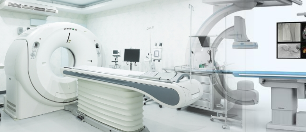
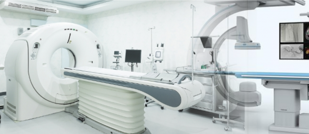
COMPUTED TOMOGRAPHY
Computed tomography is a radiological diagnostic method aimed at creating a cross-sectional image of the examined area of the body using x-ray. CT has made it possible to evaluate many parts of the body that cannot be shown by direct radiographs, such as the brain. In addition, it has enabled the diagnosis of many diseases earlier and more accurately than other imaging methods. CT examinations help doctors in the differential diagnosis of a simple cyst and a solid tumor (a mass of tissue formed due to the rapid proliferation of some cells, ur), which provides a better assessment of diseases.
MAMMOGRAPHY
Mammography is a device used to detect early diagnosis of breast diseases, especially breast cancer. Mammography is the removal of a detailed image of breast tissue with the help of a special x-ray with a low dosage. Mammography uses low-dose x-ray, high-contrast and high-density films, as well as specially designed X-ray devices.
Early detection is very important for the success of breast cancer treatment. A mammogram can display changes in the breast even when they cannot be felt by the woman herself or her doctor. Mammography for control purposes is used to diagnose possible breast cancers at an early stage in women who have no complaints. When mammography for control purposes is performed regularly, it significantly increases the chances of success of treatment by increasing the likelihood of early diagnosis. it is recommended that every woman over the age of 40 have a mammogram once a year for control purposes.
If the first shot is normal for postmenopausal women with a high risk of bone loss, control shots are recommended every 3 years. If bone loss is involved, the control period should be determined by the treating physician.
Ultrasonography (USG) (US-Ultrasonography)
Ultrasound is a diagnostic method that displays internal organs using sound waves of such a high frequency that the human ear cannot hear. Radiation is not used in ultrasound, so it can be easily used in pregnant women and babies. Ultrasound is mainly used for imaging of intra-abdominal organs such as the liver, gallbladder, pancreas, spleen, kidneys, bladder, ovaries, and uterus.
The most common complaint of patients applying for ultrasound is abdominal pain. The growth of intra-abdominal organs such as the liver and spleen, gallbladder and kidney stones, appendicitis, ovarian cysts, and tumors in the abdomen are some of the diseases that can be diagnosed by ultrasound. In addition, superficial tissue examinations and sonographic examination of other parts can be performed.
Doppler ultrasonography
With the help of Doppler Ultrasound, one can study the presence of blood flow in an organ or its vessel, the form of current, constrictions and blockages in the vessels. The amount of blood flow, the presence of a structure that blocks the flow, whether the current is in the normal direction can be assessed. Doppler examinations are performed with the usual ultrasound devices. However, different computer equipment is available in them.
Doppler Ultrasound examination areas
– Veins of the arms and legs,
– Vessels supplying the liver,
– The vessels that supply the kidney,
– Vessels of the neck,
– Vessels belonging to the mother and fetus in pregnant women,
– Veins that feed the testicles in men.
Bone densitometry (Bone density device)
A bone densitometry is a test conducted to evaluate bone density loss for screening and measuring bone mineral density. This test is used to diagnose osteoporosis, a bone condition that develops as a result of calcium loss in the bones.
– Bone densitometry is a standard method for assessing bone mineral density.
– Provides quick and painless measurement of bone loss.
OUR DOCTORS